GE VIVID S60
Lorem ipsum dolor sit amet, consectetur adipiscing elit. Fusce vitae dolor ipsum. Sed nec euismod metus. Donec lacinia lorem sapien, et accumsan massa auctor ac. Ut eget lacus vel sapien pretium viverra ut at elit. Ut id nulla lacus. Donec nec faucibus sapien. Praesent id purus sit amet neque ornare mollis vulputate sed augue. Aliquam erat volutpat. Donec purus justo, lacinia eu nunc et, ornare maximus urna.
Curabitur quis magna nisi. Praesent finibus magna consequat, tempus massa non, cursus nisl. Curabitur eget diam vel ligula venenatis maximus. Integer faucibus dignissim ex at interdum. Nam mattis massa nec suscipit elementum. Ut quis iaculis nisi. Donec bibendum cursus metus, id finibus erat congue in. Aenean mi magna, varius a condimentum sed, egestas id mi. Mauris gravida orci at odio scelerisque gravida. Cras vitae nisi sodales, porttitor tellus non, ornare velit. Ut neque velit, fermentum non nulla eu, porttitor luctus ligula. Aliquam ut facilisis lectus. Nullam at hendrerit erat.
• Width: 54 cm, 21.4¨
• Depth: 76 cm, 30.2¨
• Height: 138 cm – 168 cm, 54.4¨ – 66.7¨
• Minimum height with folded screen: 112 cm, 44.4¨
• Weight: <73 kg, 161 lbs
GE Vivid S60 Specifications:
• Infinite number of effective channels
• Minimum field-of-view range (depth): 0 – 2 cm (zoom) (probe dependent)
• Maximum field-of-view range (depth): 0 – 36 cm (probe dependent)
• Width range: 10 – 120 degrees
• Continuous dynamic receive focus/ continuous dynamic receive aperture
• Continuous dynamic transmit focus
• Adjustable dynamic range, infinite upper level
GE Vivid S60 Electrical power:
• Nominal input voltage: 100-240 VAC, 50/60 Hz
• Rated power consumption: 500 VA
The GE Vivid S70 and the GE Vivid S60 are GE’s new cardiovascular systems who cover a wide range of price points from midrange to high-end cardiac, effectively meeting the requirements of long-term GE Vivid S6 and GE Vivid S5 customers.
The GE Vivid S70 and the GE Vivid S60 are empowered by GE’s new cSound beamformer software and image reconstruction platform. However, the GE Vivid S60 does not support XDClear transducers like the GE Vivid S70 or the Vivid E series. The major difference with the GE Vivid S60 is that the GE Vivid S70 supports 4D TEE and XDclear technology.
The GE Vivid S60 is a good option for physicians considering to have high-end 2D and Doppler image quality without premium features. Its 19¨ monitor seems a bit small when compared with those of competitors, such as the 21.5” wide LCD monitor from the Affiniti series. However, the larger 12¨ LCD touchscreen will provide support for a more comfortable and efficient workflow.
The biggest weakness of the Vivid S70 and the Vivid S60 is the narrow range of transducers; there is only one linear option: the 9L-D. A couple of linear probes and an intraoperative probe are expected to be added soon. If 4D TEE is not a reason for purchasing a high-end cardiac system, then the GE Vivid S60, or a factory-refurbished GE Vivid E9 can be also be a great option to consider.
Cardiac Sector Probe
3Sc-RS[1.3 – 4.5MHz]
6S-D [2.4 – 8.0MHz]
12S-D [4.0 – 12.0MHz
Linear Probe
9L-D [2.4 – 10.0MHz]
Convex Probe
C1-6-D [1.5 – 6.0MHz]
Non-imaging Doppler Probe
P2D [2 MHz]
Audult TEE Probe
6Tc-RS [3.0 – 8.0MHz]
Pediatric TEE Probe
9T-RS [3.0 – 10.0MHz]
Volume TEE Probe
6VT-D [3.0 – 8.0MHz]
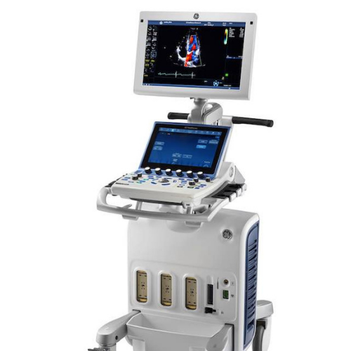
• 19¨ wide screen High-Definition (HD) flicker-free LCD display
• 12¨ ultra-high-resolution, multi-touch LCD wide screen
• 4 active probe ports and one CW port
• ECG port
• Integrated HDD
• Multiple USB ports (front/back)
• Integrated DVD-R multi drive (optional )
• On-board storage for B/W thermal printer
• Integrated speakers for premium sound
• Four swivel wheels – front wheel breaks, rear wheels direction lock
• Integrated cable management
• Easily accessible removable air filters for cleaning
• Front and rear handles
• Side storage trays
• Hand rest
• Automatic Optimization
• CrossXBeam
• Speckle Reduction Imaging (SRI-HD)
• Coded phase inversion
• Tissue velocity M-mode
• Continuous wave Doppler
• Tissue M-mode
• Pulsed wave Doppler
• Anatomical M-mode
• Curved anatomical M-mode
• Tissue velocity imaging
• Tissue velocity Doppler
• B-flow
• Raw Data Analysis
• Real-time automatic Doppler calcs
• Tissue Tracking Mode
• Extended field-of-view (LOGIQView)
• LVO Contrast
• Sony Digital UP-D711 Termal Printer
• Sony Fixture Kit for Digital UP-D711 Thermal Printer
• Sony Digital UP-D25 Color Thermal Printer
• Sony Digital UP-D897 BW Thermal Printer
• Mitsubishi P93W/E Thermal Printer
• Mitsubishi P95DW Thermal Printer
• USB B/W video printer with control from system
• External USB printer connection
• 16 GB encrypted memory stick
• Three-pedal configurable footswitch
• DVI-I
• Ethernet – 10 Mbps, 100 Mbps, 1 Gbps
• Multiple USB 2.0 ports
• RS-DLP Probe Adapter for TEE Probe
• Integrated gel holders
• ECG + AHA/IEC® Cables
GE Vivid S60 Supplies:
• Aquasonic ultrasound gel
• Sono ultrasound wipes
• Sony UPP-110HG thermal printing paper
• Sony UPC-21L color thermal printing pack
• Mitsubishi KP95HG thermal roll paper (new)
• Mitsubishi KP65HM-CE High density thermal paper
GE Vivid S60 ports:
• DVI-I
• Ethernet – 10 Mbps, 100 Mbps, 1 Gbps
• Multiple USB 2.0 ports
GE Vivid S60 image storage:
• On-board database of patient information from past exams
• Storage formats:
– DICOM®-compressed or uncompressed, single/multi-frame, with/without raw data, storage via clipboard and/or seamlessly directly to destination device
– Transfer/ “Save As” JPEG, MPEG, AVI, DICOM, Raw DICOM and VolDicom formats
• Storage devices:
– USB memory stick: 16 GB
– CD-RW storage: 700 MB (DVD option required)
– DVD storage: -R (4.7 GB) (DVD option required)
– Hard drive image storage: 0.5 TB
• Compare old images with current exam
• Reload of archived data sets
• Activation control of USB devices
• Tissue synchronization imaging
• Intima Media Thickness (IMT)
• CW Doppler
• Strain/Strain Rate Mode (option, enabled by Advanced QScan)
• Tissue Synchronization Imaging Mode (option, enabled by Advanced QScan)
• B-flow
• 2D stress
• Auto EF
• Automated Function Imaging (AFI)
• Automated Ejection-Fraction Calculation (AutoEF)
• Contrast Low MI(Research only)
• Z-Scores
• Quantitative Analysis Package (Q-Analysis)
• EchoPAC™/Patient Archive
• Insite™ Express Connection (ExC) Enables Remote Service and Training
• Cardiac
• Stress (optional )
• Abdominal
• Peripheral vascular
• Fetal heart
• Pediatrics
• Neonatal cephalic
• Adult cephalic
• Small parts
• Thyroid
• Musculoskeletal
• Urology
• Rodent (optional )
• Transesophageal
• OB/GYN
• Coronary
• LVO contrast (optional )
• Nerves